Radius Bone Labelled Diagram : 12 photos of the labelled diagram of radius bone.. Each bone is a complex living organ that is made up of many cells, protein fibers, and minerals. The radius bone is the lateral bone of the forearm, and is homologous with the tibia of the lower limb. The charsi of medical literature. I'm not sure of what you mean by bone diagram. All land vertebrates have this bone.
Each bone is a complex living organ that is made up of many cells, protein fibers, and minerals. The radius is the bone which is present laterally, which means when your palm is facing upwards, it is away from the middle of your body. Labeled radius anterior view left radius coxal bone unlabeled long bone diagram unlabeled radius bone landmarks radius radial tuberosity neck of radius bone radius vs ulna bone ulna bone location radius and ulna atlas radius bone features tibia fibula radius ulna where is the. The radius bone is this bone here and it lies laterally in the anatomical position. Diagram of wrist have bone fracture.
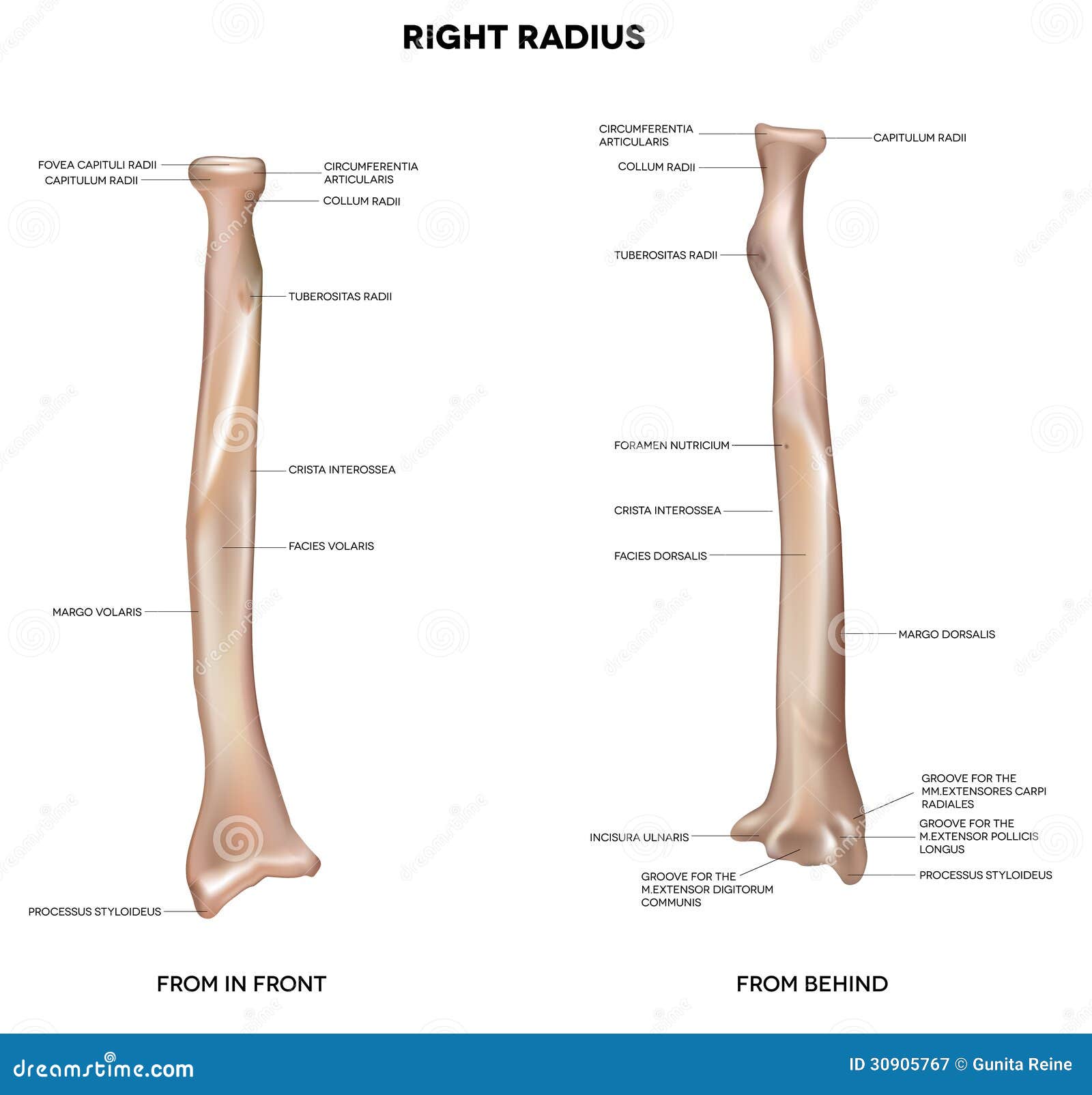
Human right radius, bone stock vector. Illustration of ... from thumbs.dreamstime.com Proximal radius (head, neck and tuberosity). Correctly label the following anatomical parts of.,label the long bone skull, clavicle, mandible, scapula, thorax, sternum, humerus, ulna, radius, carpus, phalanges (fingers), metacarpus, spine, pelvis, sacrum, femur, tibia. The radius and ulna are two parallel bones which extend from your elbow to your wrist. Illustration with skeleton of human hand ulna, radius and humerus bones. (ii) name the structure labelled a, which attaches muscle to bone. Radius along with ulna connects elbow to forearm. At the humerus, they articulate with the condyle. Learn everything about the anatomy of radius and ulna with our articles, video tutorials, labeled diagrams, and quizzes.
Labeled anatomy chart with two bones, articular cartilage, joint cavity, synovial fluid, muscle and tendon.
The radius bone is a long horizontal bone present in the forearm and is also called the radial bone. Learn everything about the anatomy of radius and ulna with our articles, video tutorials, labeled diagrams, and quizzes. It extends obliquely downward into a strong, conical projection. The radius and ulna are the two bones of the forearm. Labeled anatomy chart with two bones, articular cartilage, joint cavity, synovial fluid, muscle and tendon. Each bone is a complex living organ that is made up of many cells, protein fibers, and minerals. Bones arm hand wrist anatomical anatomy and carpal forearm illustration labels phalanges phalanx radius ray skeleton ulna x. Radius, in anatomy, the outer of the two bones of the forearm when viewed with the palm facing forward. The photograph may be purchased as wall art, home decor, apparel, phone cases, greeting cards, and more. It extends from the lateral side of the elbow to the thumb side of the wrist and runs parallel to the ulna. The lateral side projects distally as the styloid process. It lies laterally and parallel to ulna, the second of the forearm bones. Learn vocabulary, terms and more with flashcards, games and other study tools.
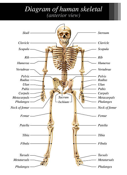
Proximal radius (head, neck and tuberosity). Forearm and hand bones labeled diagram. Lower jaw (mandible) collar bone. The radius and ulna together constitute the forearm. Left human arm is designed based on original size of relevant human bones.
The posterior and anterior views of the humeral bone ... from i.pinimg.com Labeling parts of a long bone ch 11 a p1 diagram quizlet from o.quizlet.com. The radius is considered the most commonly fractured bone in the human body, with distal radius fractures being the most common form of radial. This ulnar view labelled illustration is from 'asklepios atlas of the human anatomy'. It extends obliquely downward into a strong, conical projection. The radius is a long bone in the forearm. Diagram of wrist have bone fracture. In its distal part, the radial shaft expands to form a rectangular end. Learn everything about the anatomy of radius and ulna with our articles, video tutorials, labeled diagrams, and quizzes.
(vi) draw a labelled diagram of the cells as seen at high magnifi cation.
A basic human skeleton is studied in schools with a simple diagram. The radius and ulna are the two bones of the forearm. The bones shown in the chest and hip region in the labeled human skeleton diagram are the ribs humerus is located in the upper arm. Learn everything about the anatomy of radius and ulna with our articles, video tutorials, labeled diagrams, and quizzes. In its distal part, the radial shaft expands to form a rectangular end. The ulna articulates with the trochlea and the radius articulates with the capitulum. The charsi of medical literature. The radius or radial bone is one of the two large bones of the forearm, the other being the ulna. A the given diagram is anterior view of…view the full answer. It extends obliquely downward into a strong, conical projection. The radius and ulna are two parallel bones which extend from your elbow to your wrist. The skeleton acts as a scaffold by providing support and protection for the soft tissues that make up the rest of the body. Cheek bone (zygoma) upper jaw (maxilla).
The radius or radial bone is one of the two large bones of the forearm, the other being the ulna. Are these bones above from the right or left side of the body? Labeled anatomy chart with two bones, articular cartilage, joint cavity, synovial fluid, muscle and tendon. The ulna articulates with the trochlea and the radius articulates with the capitulum. (ii) name the structure labelled a, which attaches muscle to bone.
Best Arm Bones Diagram Stock Photos, Pictures & Royalty ... from media.istockphoto.com Labeled anatomy chart with two bones, articular cartilage, joint cavity, synovial fluid, muscle and tendon. Left human arm is designed based on original size of relevant human bones. (ii) name the structure labelled a, which attaches muscle to bone. Labeled radius anterior view left radius coxal bone unlabeled long bone diagram unlabeled radius bone landmarks radius radial tuberosity neck of radius bone radius vs ulna bone ulna bone location radius and ulna atlas radius bone features tibia fibula radius ulna where is the. In humans it is shorter than the other bone of the forearm, the ulna. The bones shown in the chest and hip region in the labeled human skeleton diagram are the ribs humerus is located in the upper arm. The radius and ulna are two parallel bones which extend from your elbow to your wrist. All land vertebrates have this bone.
The radius is the lateral bone of the forearm, and is homologous with the tibia of the lower limb.
The skeleton acts as a scaffold by providing support and protection for the soft tissues that make up the rest of the body. (ii) name the structure labelled a, which attaches muscle to bone. Individually selectable every part, ideal for learning. The radius or radial bone is one of the two large bones of the forearm, the other being the ulna. It lies laterally and parallel to ulna, the second of the forearm bones. Labeled radius anterior view left radius coxal bone unlabeled long bone diagram unlabeled radius bone landmarks radius radial tuberosity neck of radius bone radius vs ulna bone ulna bone location radius and ulna atlas radius bone features tibia fibula radius ulna where is the. The radius is considered the most commonly fractured bone in the human body, with distal radius fractures being the most common form of radial. The radius bone is shorter. The radius is the lateral bone of the forearm, and is homologous with the tibia of the lower limb. The radius bone (os radius) supports the lateral (thumb) side of the forearm and the ulna bone (os ulna) supports the medial (little finger) side. The radius is a long bone in the forearm. It is simulated by using a 12 kg/cm servo motor with gears. Radius along with ulna connects elbow to forearm.
In the medial surface, there labelled radius bone. It extends from the lateral side of the elbow to the thumb the upper extremity of the radius consists of a somewhat cylindrical head articulating with the ulna and the humerus, a neck, and a radial tuberosity.


0 Komentar